Formulation Supplied at 0.5 mg/ml in Tris saline, 0.02% sodium azide, pH7.3 with 0.5% bovine serum albumin.
| |
Unit Size 100 µg | |
Storage Instructions Aliquot and store at -20°C. Minimize freezing and thawing. | |
Synonym / Alias Names ANGPT1|angiopoietin 1|ANG1|AGPT|AGP1 | |
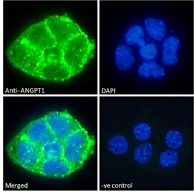
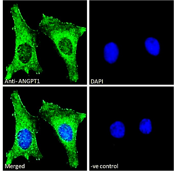
Usage Summary Immunofluorescence: Strong expression of the protein seen in the plasma membranes of A431 and HeLa cells. Recommended concentration: 10µg/ml.
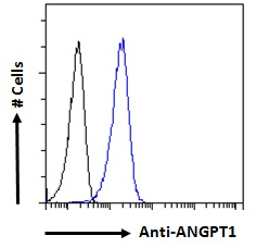
Flow Cytometry: Flow cytometric analysis of A431 cells. Recommended concentration: 10ug/ml. | |
Accession ID NP_001137.2 | |
Blocking Peptide EBP10272 | |
Immunogen Peptide with sequence C-PDFSSQKLQH, from the internal region of the protein sequence according to NP_001137.2. | |
Peptide Sequence C-PDFSSQKLQH | |
Purification Method Purified from goat serum by ammonium sulphate precipitation followed by antigen affinity chromatography using the immunizing peptide. | |
Shipping Instructions Refrigerated | |
Predicted Species Human, Mouse, Rat, Dog, Cow, Pig | |
Reactive Species Human | |
Human Gene ID 284 | |
Mouse Gene ID 11600 | |
Rat Gene ID 89807 | |
Product Grade  | |
ELISA Detection Limit Antibody detection limit dilution 1:32000. | |
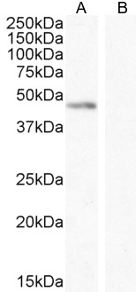
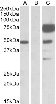
Western Blot In transfected HEK293 transiently expressing full-length Human ANGPT1 (myc and DYKDDDDK tagged), a band of approx. 48kDa was observed. No bands were observed in mock-transfected HEK293 and the 48kDa plus a 75kDa band was observed using anti-DYKDDDDK tag antibody. An approx. 48kDa band was also observed in lysates of cell line K562 and was successfully blocked by incubation with the immunizing peptide. Recommended concentration: 0.1-1µg/ml. Primary incubation 1 hour at room temperature. | |
Application Type Pep-ELISA, WB, WB-Trf, IF, FC |
Goat Anti-ANGPT1 Antibody
$431.00
| SKU | Unit Size | Price |
|---|---|---|
Select a unit size:
Documents |