Formulation Supplied at 0.5 mg/ml in Tris saline, 0.02% sodium azide, pH7.3 with 0.5% bovine serum albumin.
| |
Unit Size 100 µg | |
Storage Instructions Aliquot and store at -20°C. Minimize freezing and thawing. | |
Synonym / Alias Names MGC142296|MGC142294|CD274 molecule|CD274|HGNC:17635|PDL1|programmed cell death 1 ligand 1|PDCD1L1|PDL1|B7H1|B7-H|PDCD1LG1|PD-L1|CD274 antigen | |
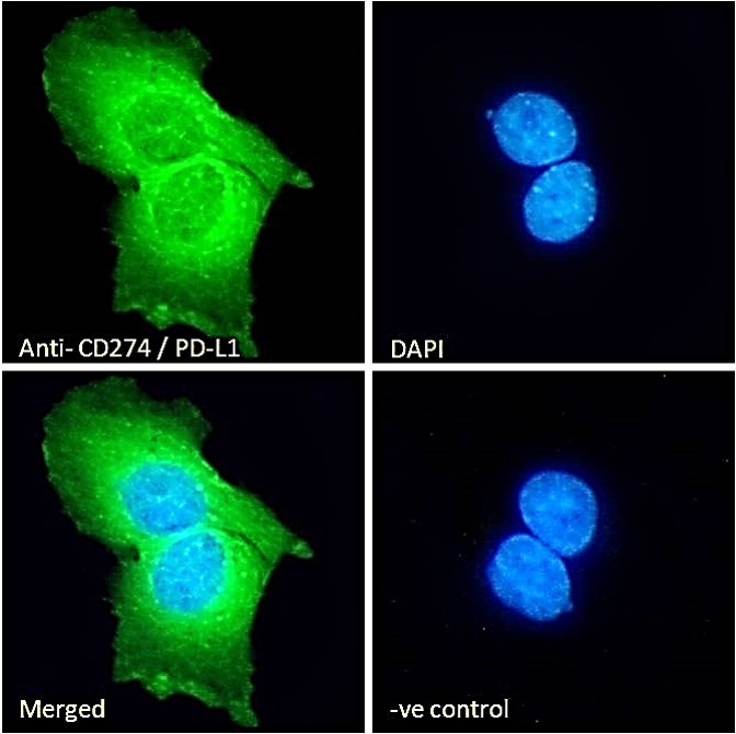
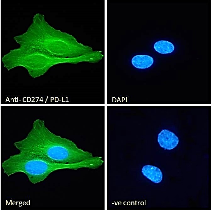
Usage Summary Immunofluorescence: Strong expression of the protein seen in th membrane and cytoplasm of U2OS and A431 cells. Recommended concentration: 10µg/ml.
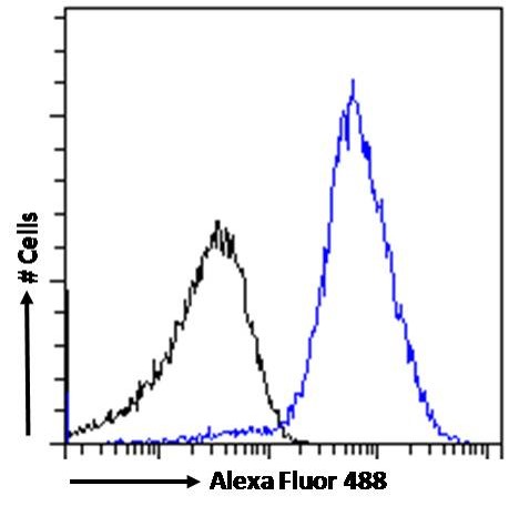
Flow Cytometry: Flow cytometric analysis of Jurkat cells. Recommended concentration: 10ug/ml. | |
Accession ID NP_054862.1; NP_001254635.1 | |
Blocking Peptide EBP06621 | |
Immunogen Peptide with sequence CKKQSDTHLEET, from the C Terminus of the protein sequence according to NP_054862.1; NP_001254635.1. | |
Product Comments This antibody is expected to recognize reported isoforms a and b (NP_054862.1; NP_001254635.1) only. | |
Peptide Sequence CKKQSDTHLEET | |
Purification Method Purified from goat serum by ammonium sulphate precipitation followed by antigen affinity chromatography using the immunizing peptide. | |
Shipping Instructions Refrigerated | |
Predicted Species Human | |
Reactive Species Human | |
Human Gene ID 29126 | |
Product Grade  | |
ELISA Detection Limit Antibody detection limit dilution 1:128000. | |
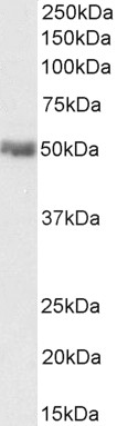
Western Blot Approx 50kDa band observed in Human Heart lysates and in lysates of cell line U2OS (calculated MW of 33.3kDa according to 1NP_054862.1).This band was successfully blocked by incubation with the immunizing peptide and is routinely observed by other sources. Recommended concentration: 0.01-0.1µg/ml. Primary incubation 1 hour at room temperature. | |
Application Type Pep-ELISA, WB, IF, FC |

Goat Anti-CD274 / PD-L1 Antibody
$431.00
| SKU | Unit Size | Price |
|---|---|---|
Select a unit size:
Selected References [{"pmid": 28026044, "intro": "This antibody (previous batch) has been successfully used in Western blot on Human:", "title": "Expression of programmed cell death-1 and its ligand B7 homolog 1 in peripheral blood lymphocytes from patients with peripartum cardiomyopathy.", "author": "Guozhi Xia, Xiaopu Zheng, Xinye Yao, Xiaowei Yao, Zhongwei Liu, Junkui Wang.", "journal": "Clin Cardiol. 2016 Dec 27."}, {"pmid": 21853301, "intro": "This antibody (previous batch) has been successfully used in Western blot and IHC on Human:", "title": "Upregulation of B7-H1 expression is associated with macrophage infiltration in hepatocellular carcinomas.", "author": "Chen J, Li G, Meng H, Fan Y, Song Y, Wang S, Zhu F, Guo C, Zhang L, Shi Y.", "journal": "Cancer Immunol Immunother. 2012 Jan;61(1):101-8."}] |
Documents |