Formulation Supplied at 0.5 mg/ml in Tris saline, 0.02% sodium azide, pH7.3 with 0.5% bovine serum albumin.
| |
Unit Size 100 µg | |
Storage Instructions Aliquot and store at -20°C. Minimize freezing and thawing. | |
Synonym / Alias Names HIES|MGC16063|FLJ20882|acute-phase response factor|DNA-binding protein APRF|APRF|signal transducer and activator of transcription 3 (acute-phase response factor)|STAT3 | |
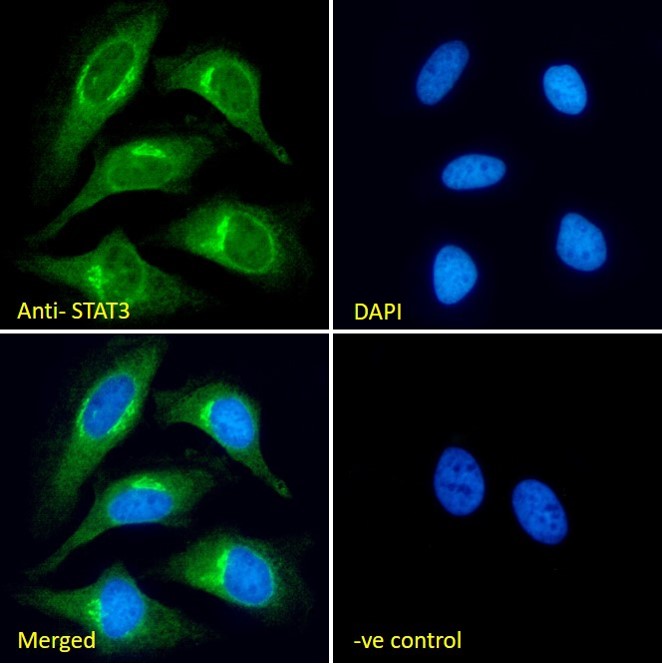
Usage Summary Immunofluorescence: Strong expression of the protein seen in the Golgi and cytoplasm of HeLa and A431 cells. Recommen
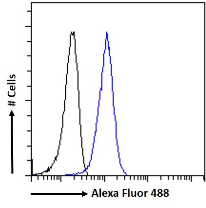
Flow Cytometry: Flow cytometric analysis of A431 cells.. Recommended concentration: 10ug/ml. ded concentration: 10µg/ml. | |
Accession ID NP_003141.2; NP_644805.1 | |
Blocking Peptide EBP05073 | |
Immunogen Peptide with sequence DMELTSECATSPM, from the C Terminus of the protein sequence according to NP_003141.2; NP_644805.1. | |
Product Comments This antibody is expected to recognise isoforms 1 and 2 (as represented by NP_644805.1 and NP_003141.2 respectively) | |
Peptide Sequence DMELTSECATSPM | |
Purification Method Purified from goat serum by ammonium sulphate precipitation followed by antigen affinity chromatography using the immunizing peptide. | |
Shipping Instructions Refrigerated | |
Predicted Species Human, Mouse, Rat, Cow | |
Reactive Species Human | |
Human Gene ID 6774 | |
Mouse Gene ID 20848 | |
Rat Gene ID 25125 | |
Product Grade  | |
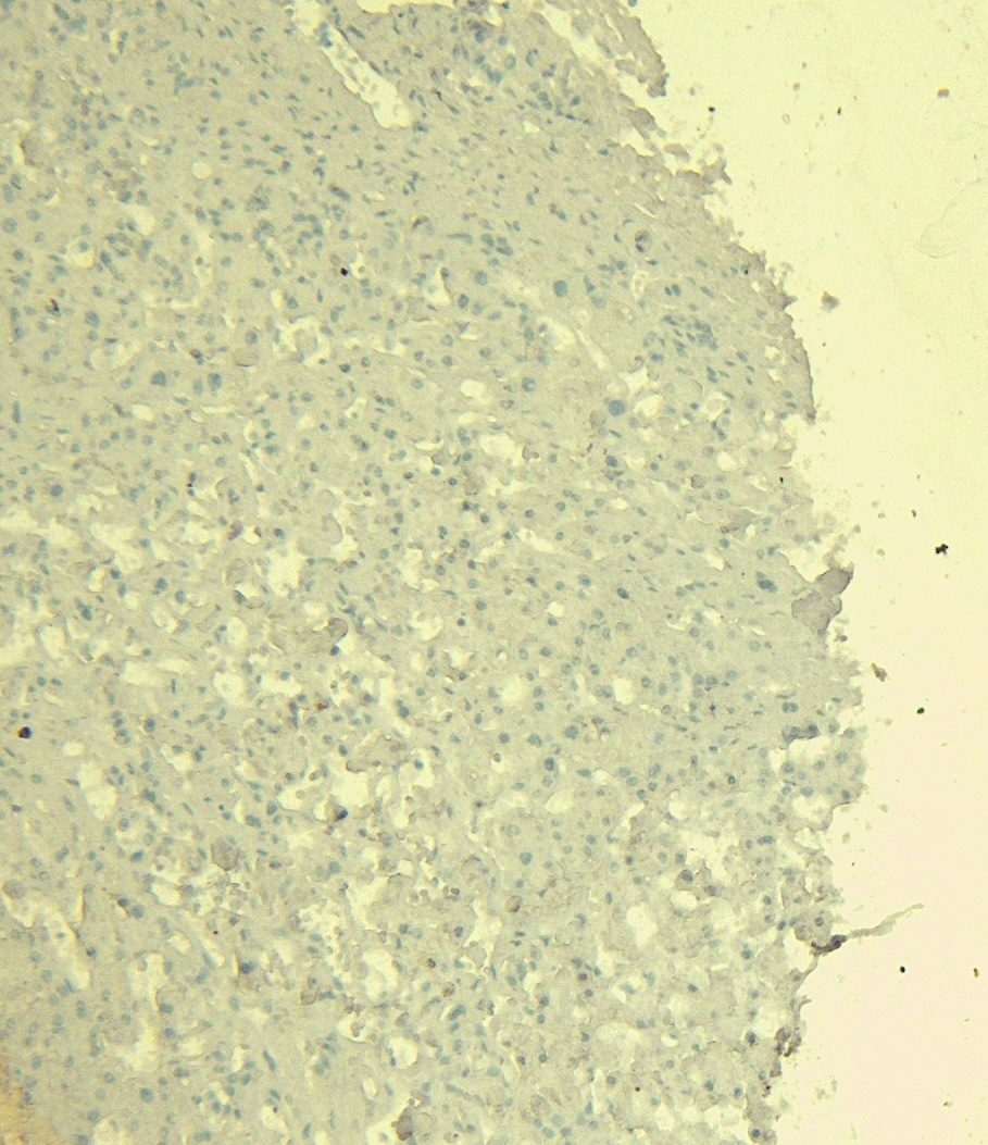
IHC Results Paraffin embedded Human Liver. Recommended concentration: 6-7µg/ml. | |
ELISA Detection Limit Antibody detection limit dilution 1:16000. | |
Application Type Pep-ELISA, IHC, IF, FC |


Goat Anti-STAT3 (isoform 1 and 2) Antibody
$431.00
| SKU | Unit Size | Price |
|---|---|---|
Select a unit size:
Documents |