Formulation Supplied at 0.5 mg/ml in Tris saline, 0.02% sodium azide, pH7.3 with 0.5% bovine serum albumin.
| |
Unit Size 100 µg | |
Storage Instructions Aliquot and store at -20°C. Minimize freezing and thawing. | |
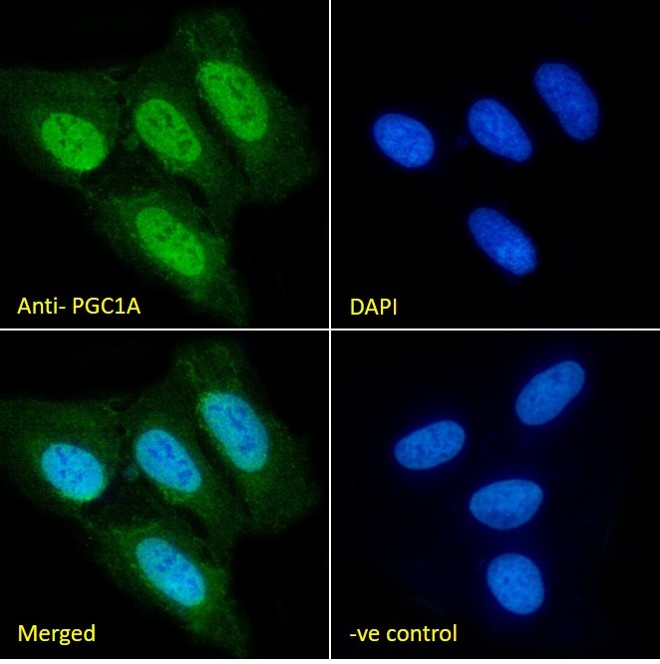
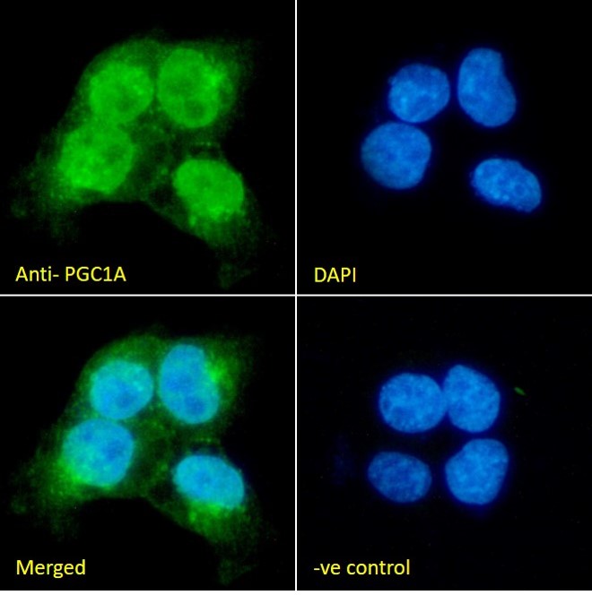
Usage Summary Immunofluorescence: Strong expression of the protein seen in A431 and U2OS cells. Recommended concentration: 10µg/ml.
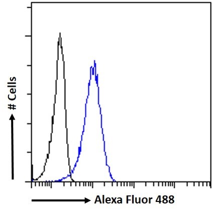
Flow Cytometry: Flow cytometric analysis of A431 cells. Recommended concentration: 10ug/ml. | |
Accession ID NP_037393.1, NP_001317680.1, NP_001317681.1, NP_001317682.1 , NP_001341756.1 | |
Blocking Peptide EBP13106 | |
Immunogen Peptide with sequence RDSVSPPKSLFSQC, from the internal region of the protein sequence according to NP_037393.1, NP_001317680.1, NP_001317681.1, NP_001317682.1 , NP_001341756.1. | |
Product Comments This antibody appears to recognise multiple isoforms and is an alternative product to EB07856. | |
Peptide Sequence RDSVSPPKSLFSQC | |
Purification Method Purified from goat serum by ammonium sulphate precipitation followed by antigen affinity chromatography using the immunizing peptide. | |
Shipping Instructions Refrigerated | |
Predicted Species Human, Mouse, Rat | |
Reactive Species Human | |
Human Gene ID 10891 | |
Product Grade  | |
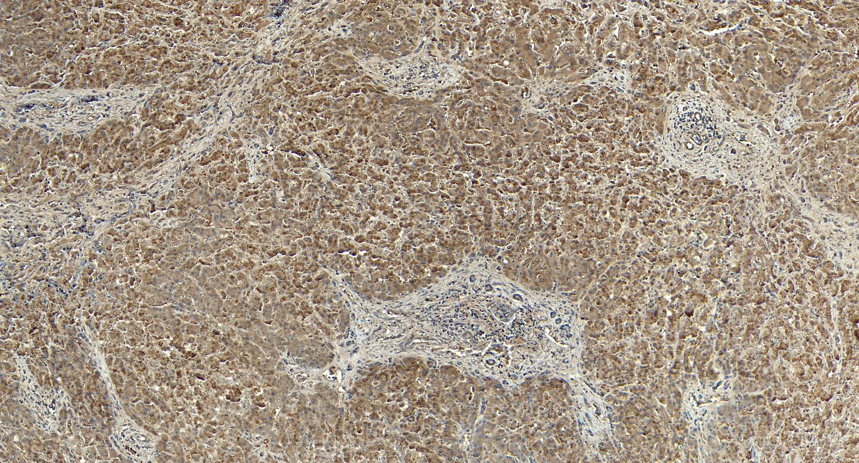
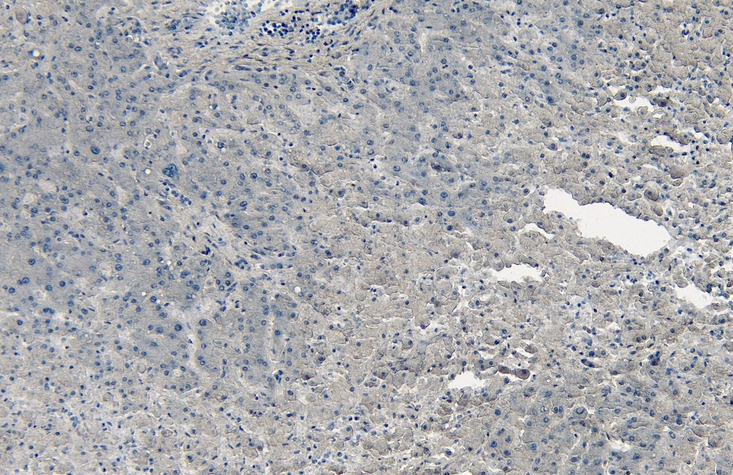
IHC Results Paraffin embedded Human Liver and Kidney. Recommended concentration: 5-6µg/ml. | |
ELISA Detection Limit Antibody detection limit dilution 1:128000. | |
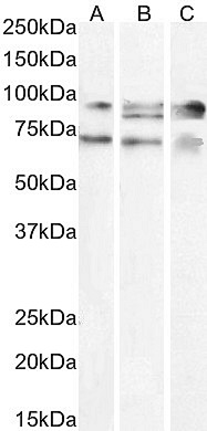
Western Blot Approx. 70+90kDa bands observed in lysates of cell line A431 and in nuclear HEK293 cell lysates, and an additional 85kDa band observed in HepG2 cell lysate (calculated MW of 91.0kDa according to NP_037393.1, 89.6kDa according to NP_001317681.1, and 77.1kDa according to NP_001317682.1). All bands were successfully blocked by incubation with the immunizing peptide. Recommended concentration: 1.5-3µg/ml. Primary incubation 1 hour at room temperature. | |
Application Type Pep-ELISA, WB, IF, IHC, FC |


Goat Anti-PPARGC1A Antibody
$260.00
| SKU | Unit Size | Price |
|---|---|---|
Select a unit size: