Formulation Supplied at 0.5 mg/ml in Tris saline, 0.02% sodium azide, pH7.3 with 0.5% bovine serum albumin.
| |
Unit Size 100 µg | |
Storage Instructions Aliquot and store at -20°C. Minimize freezing and thawing. | |
Synonym / Alias Names betaglycan proteoglycan|betaglycan|BGCAN|transforming growth factor, beta receptor III|TGFBR3 | |
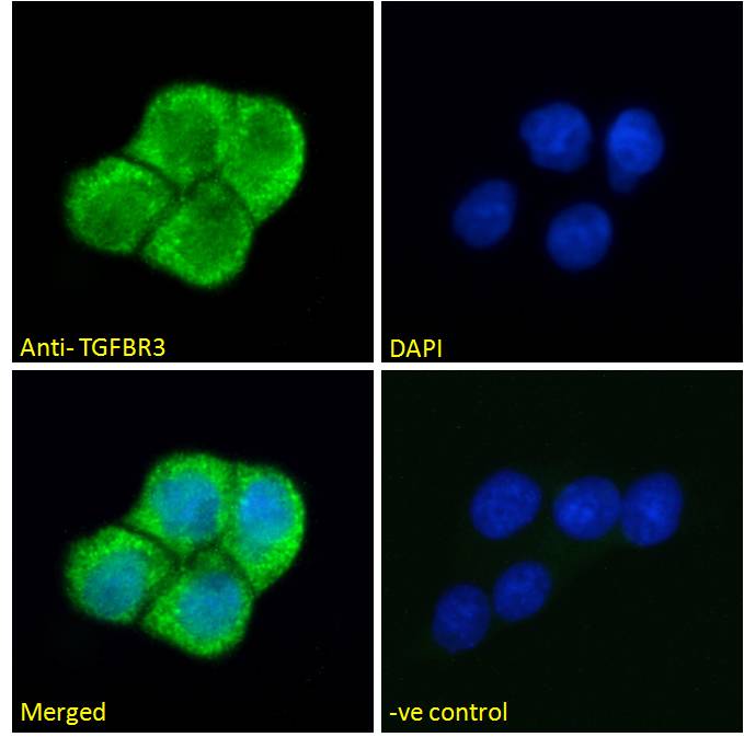
Usage Summary Immunofluorescence: Strong expression of the protein seen in the cytoplasm of A431 cells. Recommended concentration: 10µg/ml.
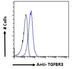
Flow Cytometry: Flow cytometric analysis of A431 cells. Recommended concentration: 10ug/ml. | |
Accession ID NP_003234.2; NP_001182613.1 | |
Blocking Peptide EBP08467 | |
Immunogen Peptide with sequence C-TKSIRDDIPSTQGN, from the internal region of the protein sequence according to NP_003234.2; NP_001182613.1. | |
Peptide Sequence C-TKSIRDDIPSTQGN | |
Purification Method Purified from goat serum by ammonium sulphate precipitation followed by antigen affinity chromatography using the immunizing peptide. | |
Shipping Instructions Refrigerated | |
Predicted Species Human | |
Reactive Species Human | |
Human Gene ID 7049 | |
Product Grade  | |
ELISA Detection Limit Antibody detection limit dilution 1:32000. | |
Western Blot Preliminary testing showed a consistent band at approx 75kDa in MCF7 and K562 cell lysates and in Human Kidney lysates after 1µg/ml antibody staining. This band was successfully blocked by incubation with the immunizing peptide. Primary incubation 1 hour at room temperature. Please note that we cannot currently find an explanation in the literature for this band, given the calculated size of 93.5kDa according to NP_003234.2). | |
Application Type Pep-ELISA, IF, FC |
Goat Anti-TGFBR3 Antibody
$431.00
| SKU | Unit Size | Price |
|---|---|---|
Select a unit size:
Documents |