
Search
Type in product code or name:
Goat Anti-PPARGC1A Antibody (EB13106)
| Code | Name | Applications | Availability | Size | Tested species | Price | Grade | |
|---|---|---|---|---|---|---|---|---|
| EB13106 | Goat Anti-PPARGC1A Antibody | Pep-ELISA, WB, IF, IHC, FC | 100µg specific antibody in 200µl | Human | $450.00 |  |
||
Ordering Information forOrder direct from Everest
$73.00: Overnight shipping (not applicable to SB-shifix) to United States of America
Sent via overnight courier arrives approximately next day to mainland USA. Please note that your order will be shipped by our US partner Abcore LLC. Order offlineNow shipping direct to US customers! 99% of products are held in stock for immediate shipment, see individual product pages for availability. Please order through the US Dollar shopping cart (links on each product page) or we accept orders by email ([email protected]), fax (888-841-9041), phone (888-320-4628). Please include the following information with your order:
In stock orders received by 13.30 PST on Monday through Thursday are shipped the same day. Orders are shipped by our US logistics partner Abcore LLC.
OR
Order from our local distributorARP American Research Products, IncARP American Research Products, Inc |
||||||||
Target Protein
Principal Names:Official Symbol: PPARGC1A
Accession Number(s): NP_037393.1, NP_001317680.1, NP_001317681.1, NP_001317682.1 , NP_001341756.1
Human GeneID(s): 10891
Important Comments: This antibody appears to recognise multiple isoforms and is an alternative product to EB07856.
Immunogen
Peptide with sequence RDSVSPPKSLFSQC, from the internal region of the protein sequence according to NP_037393.1, NP_001317680.1, NP_001317681.1, NP_001317682.1 , NP_001341756.1.
Please note the peptide is available for sale.
Purification and Storage
Purified from goat serum by ammonium sulphate precipitation followed by antigen affinity chromatography using the immunizing peptide.
Supplied at 0.5 mg/ml in Tris saline, 0.02% sodium azide, pH7.3 with 0.5% bovine serum albumin.
Aliquot and store at -20°C. Minimize freezing and thawing.
Applications Tested
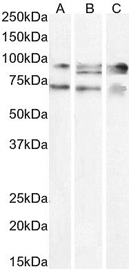
Peptide ELISA: antibody detection limit dilution 1:128000.Western blot: Approx. 70+90kDa bands observed in lysates of cell line A431 and in nuclear HEK293 cell lysates, and an additional 85kDa band observed in HepG2 cell lysate (calculated MW of 91.0kDa according to NP_037393.1, 89.6kDa according to NP_001317681.1, and 77.1kDa according to NP_001317682.1). All bands were successfully blocked by incubation with the immunizing peptide. Recommended concentration: 1.5-3µg/ml. Primary incubation 1 hour at room temperature.
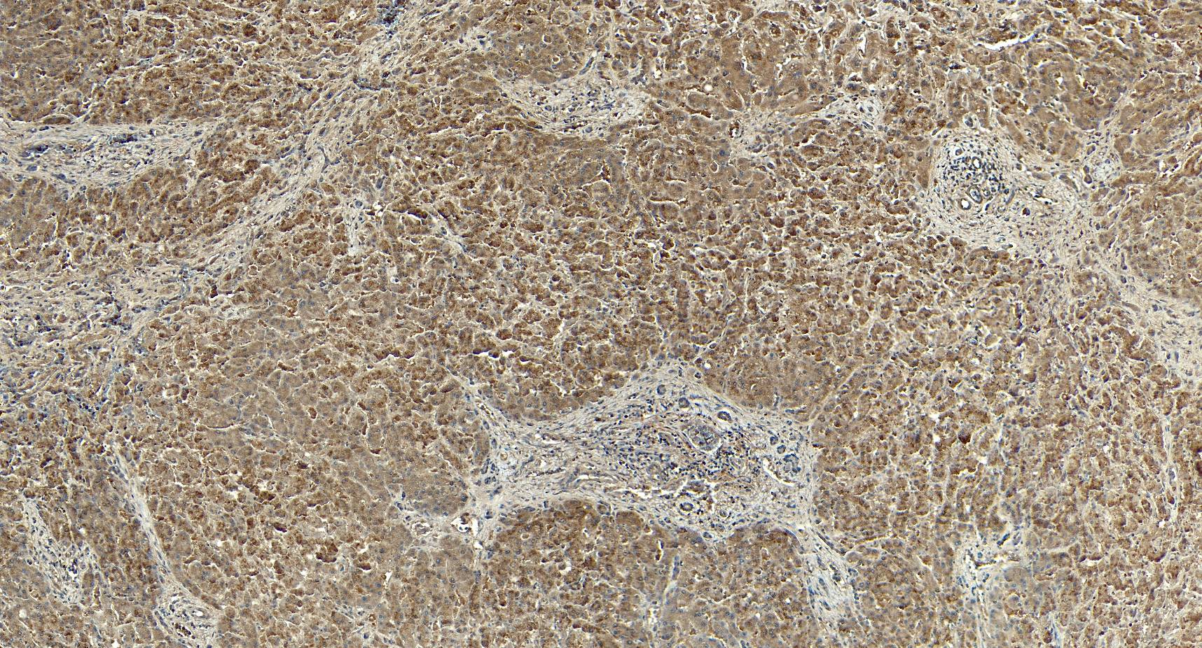
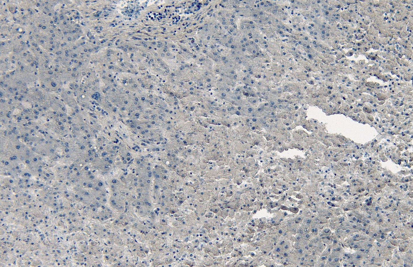
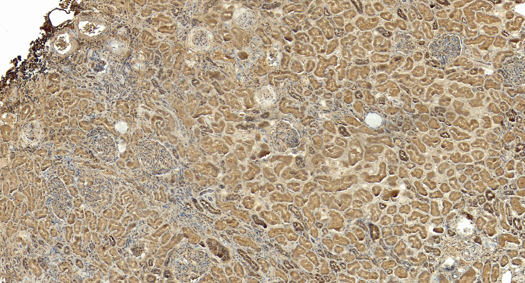
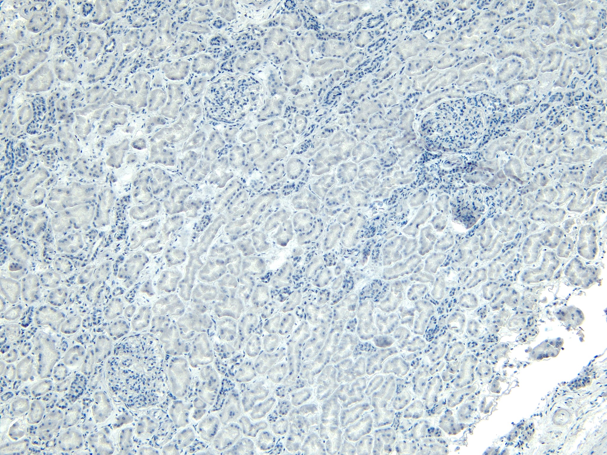
IHC: Paraffin embedded Human Liver and Kidney. Recommended concentration: 5-6µg/ml.
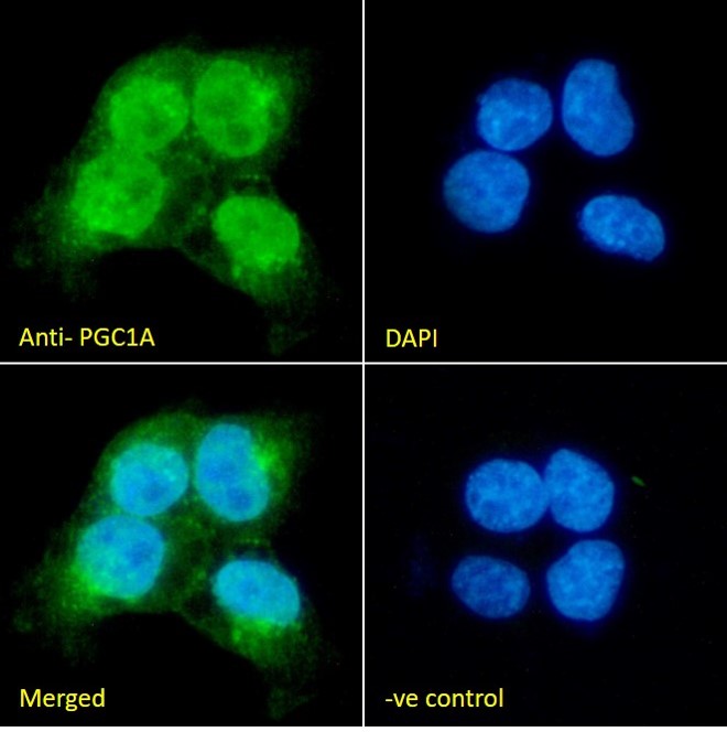
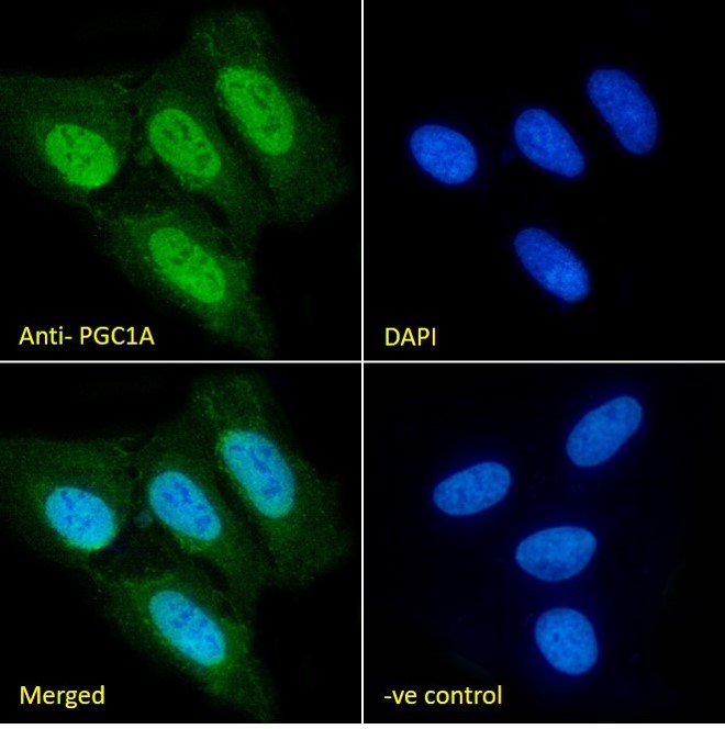
Immunofluorescence: Strong expression of the protein seen in A431 and U2OS cells. Recommended concentration: 10µg/ml.
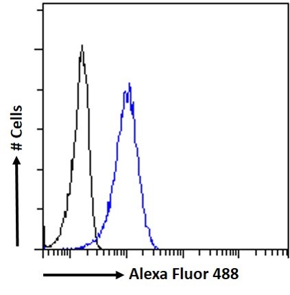
Flow Cytometry: Flow cytometric analysis of A431 cells. Recommended concentration: 10ug/ml.
Species Reactivity
Tested: HumanExpected from sequence similarity: Human, Mouse, Rat
Product Reviews
Please login to review this product.- Western blot Guidelines
- Everest Western blot labeling
- Western blot Trouble shooting guide
- IHC staining on paraffin sections
- Immunofluorescence Protocol
- Flow Cytometry Protocol
- MSDS - All Goat antibodies
- Tissue Lysate Preparation
- Cell Lysate Preparation
- Blocking with the immunizing peptide
- LsBio IHC Protocol