Formulation Supplied at 0.5 mg/ml in Tris saline, 0.02% sodium azide, pH7.3 with 0.5% bovine serum albumin.
| |
Unit Size 100 µg | |
Storage Instructions Aliquot and store at -20°C. Minimize freezing and thawing. | |
Synonym / Alias Names calgranulin B|S100 calcium-binding protein A9 (calgranulin B)|S100 calcium-binding protein A9|S100 calcium binding protein A9 (calgranulin B)|P14|NIF|MRP14|MIF|MAC387|LIAG|L1AG|CGLB|CFAG|CAGB|60B8AG|S100 calcium binding protein A9|S100A9 | |
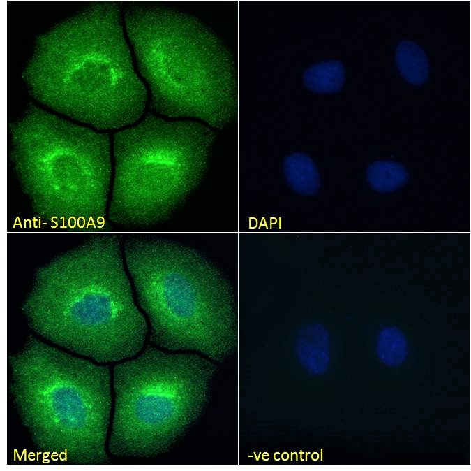
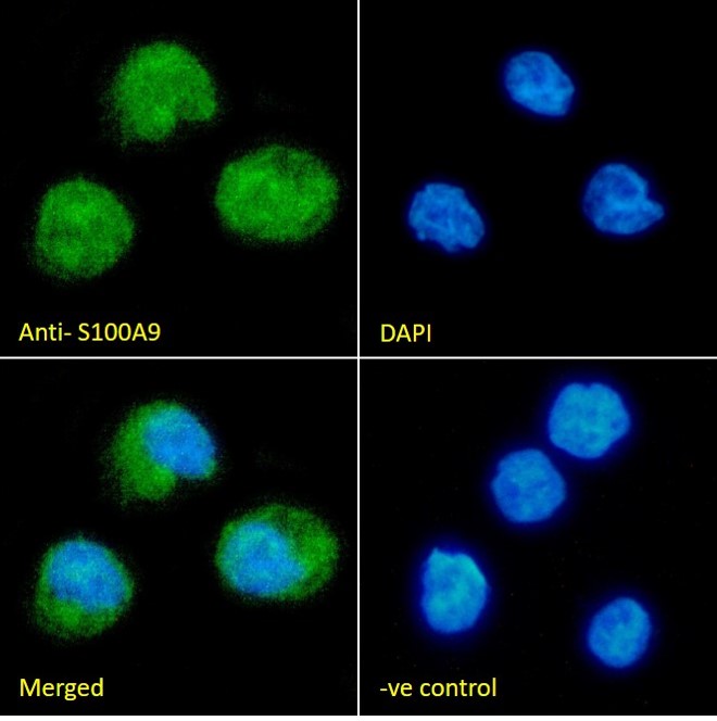
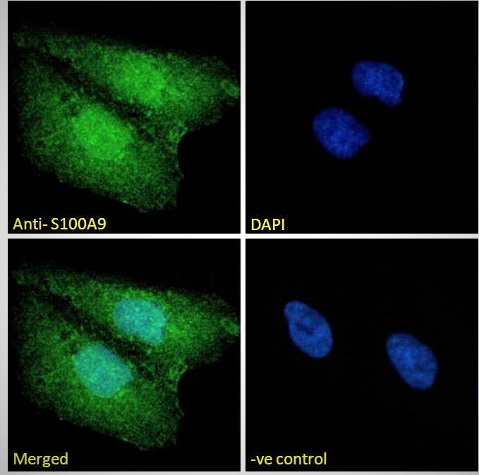
Usage Summary Immunofluorescence: Strong expression of the protein seen in the cytoplasm and nuclei of MCF7, U2OS and THP-1 cells. Recommended concentration: 10µg/ml.
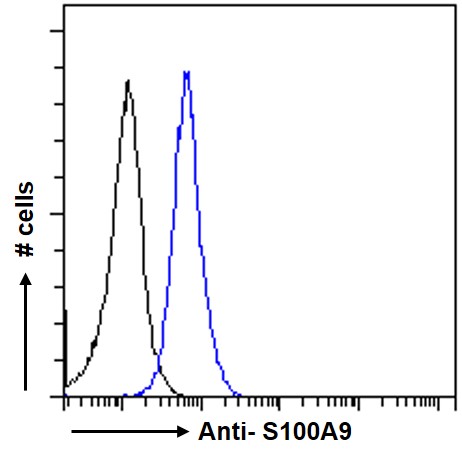
Flow Cytometry: Flow cytometric analysis of MCF7 cells. Recommended concentration: 10ug/ml. | |
Accession ID NP_002956.1 | |
Blocking Peptide EBP09146 | |
Immunogen Peptide with sequence C-DTNADKQLSFEEF , from the internal region of the protein sequence according to NP_002956.1. | |
Peptide Sequence C-DTNADKQLSFEEF | |
Purification Method Purified from goat serum by ammonium sulphate precipitation followed by antigen affinity chromatography using the immunizing peptide. | |
Shipping Instructions Refrigerated | |
Predicted Species Human | |
Reactive Species Human | |
Human Gene ID 6280 | |
Product Grade  | |
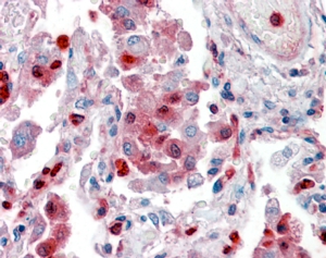
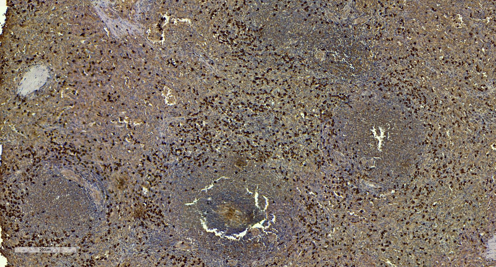
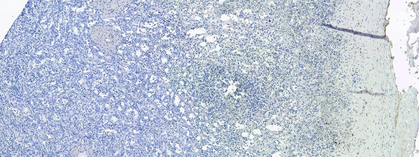
IHC Results Paraffin embedded Human Lung. Recommended concentration: 2-4µg/ml.
Paraffin embedded Human Spleen. Recommended concentration: 6-7µg/ml. | |
ELISA Detection Limit Antibody detection limit dilution 1:8000. | |
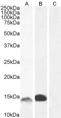
Western Blot Approx 13-14kDa band observed in Human Bone Marrow lysates and approx 14-15kDa in Human Gastrointestinal cancer lysates (calculated MW of 13.2kDa according to NP_002956.1). Recommended concentration: 0.5-2µg/ml. Primary incubation 1 hour at room temperature. | |
Application Type Pep-ELISA, WB, IHC, IF, FC |
Goat Anti-S100A9 Antibody
$431.00
| SKU | Unit Size | Price |
|---|---|---|
Select a unit size:
Documents |