
Search
Type in product code or name:
Goat Anti-AIFM1 Antibody (EB12622)
| Code | Name | Applications | Availability | Size | Tested species | Price | Grade | |
|---|---|---|---|---|---|---|---|---|
| EB12622 | Goat Anti-AIFM1 Antibody | Pep-ELISA, WB, IHC, IF | 100µg specific antibody in 200µl | Human, Mouse, Rat, Pig | $464.00 |  |
||
Ordering Information forOrder direct from EverestOrder offline99% of products are held in stock for immediate shipment, see individual product pages for availability. Please order through the US Dollar shopping cart (links on each product page) or we accept orders by email ([email protected])
In stock orders received by 11 am PST on Monday through Friday are shipped the same day. Orders are shipped by our parent company Vector Laboratories, 6737 Mowry Avenue, Newark CA 94560, USA. |
||||||||
Target Protein
Principal Names: AIFM1, apoptosis-inducing factor, mitochondrion-associated, 1, AIF, CMTX4, COWCK, COXPD6, PDCD8, apoptosis-inducing factor 1, mitochondrial, programmed cell death 8 (apoptosis-inducing factor), striatal apoptosis-inducing factorOfficial Symbol: AIFM1
Accession Number(s): NP_004199.1; NP_665811.1; NP_665812.1; NP_001124318.1
Human GeneID(s): 9131
Non-Human GeneID(s): 26926 (mouse), 83533 (rat)
Important Comments: This antibody is expected to recognize isoform 1 (NP_004199.1), isoform 2 (NP_665811.1), isoform 3 (NP_665812.1) and isoform 4 (NP_001124318.1).
Immunogen
Peptide with sequence C-NEVAKLFNIHED, from the C Terminus of the protein sequence according to NP_004199.1; NP_665811.1; NP_665812.1; NP_001124318.1.
Please note the peptide is available for sale.
Purification and Storage
Purified from goat serum by ammonium sulphate precipitation followed by antigen affinity chromatography using the immunizing peptide.
Supplied at 0.5 mg/ml in Tris saline, 0.02% sodium azide, pH7.3 with 0.5% bovine serum albumin.
Aliquot and store at -20°C. Minimize freezing and thawing.
Applications Tested
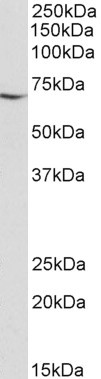
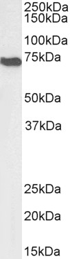
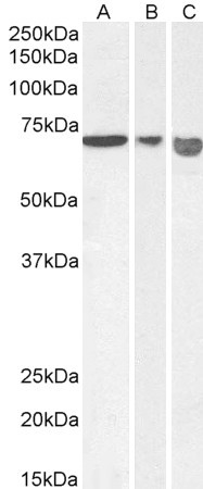
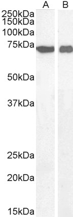
Peptide ELISA: antibody detection limit dilution 1:128000.Western blot: Approx 70kDa band observed in lysates of cell line Jurkat and NIH3T3, and in Mouse and Rat Heart and Kidney lysates. Approx. 65kDa observed in Pig Heart lysates (calculated MW of 66.9kDa according to Human NP_004199.1 and 66.8kDa according to Mouse NP_036149.1, Rat NP_112646.1 and Pig NP_001284561.1). Recommended concentration: 0.01-0.1µg/ml. Primary incubation 1 hour at room temperature.
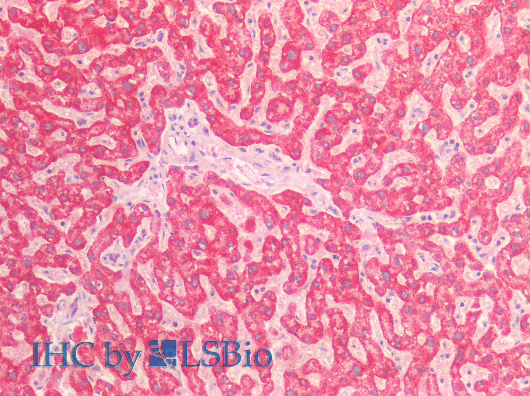
IHC: Paraffin embedded Human Liver. Recommended concentration: 5µg/ml.
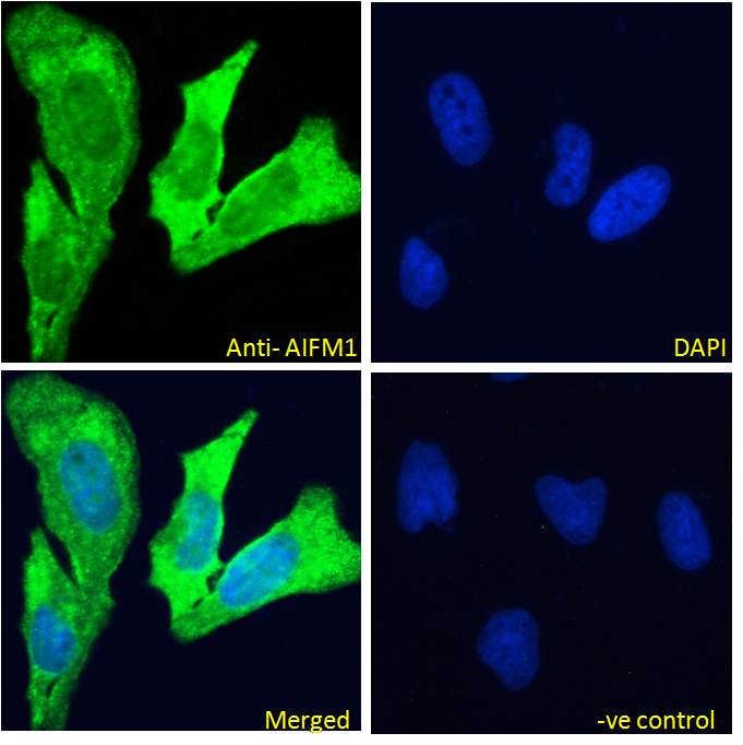
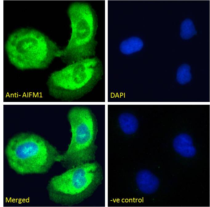
Immunofluorescence: Strong expression of the protein seen in the Mitochondria of HeLa and U2OS cells. Recommended concentration: 10µg/ml.
Species Reactivity
Tested: Human, Mouse, Rat, PigExpected from sequence similarity: Human, Mouse, Rat, Dog, Pig, Cow
Product Reviews
Please login to review this product.- Western blot Guidelines
- Everest Western blot labeling
- Western blot Trouble shooting guide
- IHC staining on paraffin sections
- Immunofluorescence Protocol
- Flow Cytometry Protocol
- MSDS - All Goat antibodies
- Tissue Lysate Preparation
- Cell Lysate Preparation
- Blocking with the immunizing peptide
- LsBio IHC Protocol