
Search
Type in product code or name:
Goat Anti-ATF6 (aa437-449) Antibody (EB12080)
| Code | Name | Applications | Availability | Size | Tested species | Price | Grade | |
|---|---|---|---|---|---|---|---|---|
| EB12080 | Goat Anti-ATF6 (aa437-449) Antibody | Pep-ELISA, WB, IF, FC | 100µg specific antibody in 200µl | Human | $464.00 |  |
||
Ordering Information forOrder direct from EverestOrder offline99% of products are held in stock for immediate shipment, see individual product pages for availability. Please order through the US Dollar shopping cart (links on each product page) or we accept orders by email ([email protected])
In stock orders received by 11 am PST on Monday through Friday are shipped the same day. Orders are shipped by our parent company Vector Laboratories, 6737 Mowry Avenue, Newark CA 94560, USA. |
||||||||
Target Protein
Principal Names: ATF6, activating transcription factor 6, ATF6A, cAMP-dependent transcription factor ATF-6 alpha, cyclic AMP-dependent transcription factor ATF-6 alphaOfficial Symbol: ATF6
Accession Number(s): NP_031374.2
Human GeneID(s): 22926
Non-Human GeneID(s): 304962 (rat)
Immunogen
Peptide with sequence C-NSYRYDHSVSNDK, from the internal region of the protein sequence according to NP_031374.2.
Please note the peptide is available for sale.
Purification and Storage
Purified from goat serum by ammonium sulphate precipitation followed by antigen affinity chromatography using the immunizing peptide.
Supplied at 0.5 mg/ml in Tris saline, 0.02% sodium azide, pH7.3 with 0.5% bovine serum albumin.
Aliquot and store at -20°C. Minimize freezing and thawing.
Applications Tested
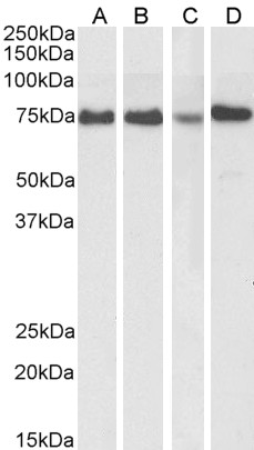
Peptide ELISA: antibody detection limit dilution 1:128000.Western blot: Approx 75-80kDa band observed in lysates of cell lines HeLa and MCF7, and in HeLa and Jurkat nuclear cell lysates (calculated MW of 74.6kDa according to NP_031374.2). Recommended concentration: 0.3-2µg/ml. Primary incubation 1 hour at room temperature.
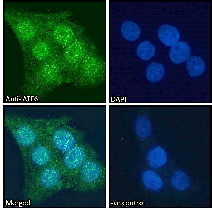
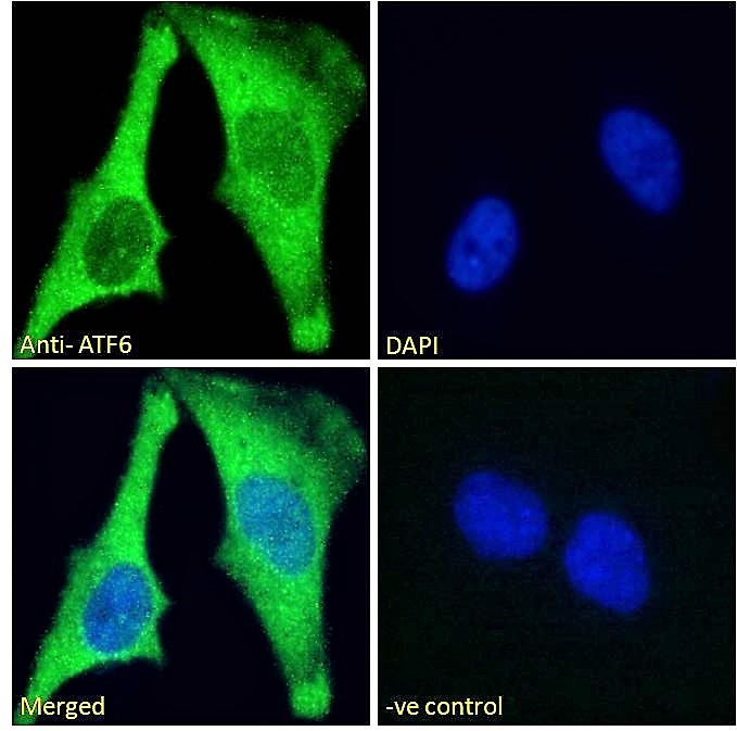
Immunofluorescence: Strong expression of the protein seen in the cytoplasm of A431 and HeLa cells. Recommended concentration: 10µg/ml.
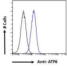
Flow Cytometry: Flow cytometric analysis of A431 cells. Recommended concentration: 10ug/ml.
Species Reactivity
Tested: HumanExpected from sequence similarity: Human, Rat, Dog, Pig, Cow
Product Reviews
Please login to review this product.- Western blot Guidelines
- Everest Western blot labeling
- Western blot Trouble shooting guide
- IHC staining on paraffin sections
- Immunofluorescence Protocol
- Flow Cytometry Protocol
- MSDS - All Goat antibodies
- Tissue Lysate Preparation
- Cell Lysate Preparation
- Blocking with the immunizing peptide
- LsBio IHC Protocol