
Search
Type in product code or name:
Goat Anti-Calcipressin-1 Antibody (EB10693)
| Code | Name | Applications | Availability | Size | Tested species | Price | Grade | |
|---|---|---|---|---|---|---|---|---|
| EB10693 | Goat Anti-Calcipressin-1 Antibody | Pep-ELISA, WB, IF, FC | 100µg specific antibody in 200µl | Human, Mouse | $464.00 |  |
||
Ordering Information forOrder direct from EverestOrder offline99% of products are held in stock for immediate shipment, see individual product pages for availability. Please order through the US Dollar shopping cart (links on each product page) or we accept orders by email ([email protected])
In stock orders received by 11 am PST on Monday through Friday are shipped the same day. Orders are shipped by our parent company Vector Laboratories, 6737 Mowry Avenue, Newark CA 94560, USA. |
||||||||
Target Protein
Principal Names: RCAN1, regulator of calcineurin 1, ADAPT78, CSP1, DSC1, DSCR1, MCIP1, RCN1, Down syndrome candidate region 1, OTTHUMP00000108621, OTTHUMP00000108622, OTTHUMP00000214669, OTTHUMP00000214670, calcipressin-1, calcium andOfficial Symbol: RCAN1
Accession Number(s): NP_004405.3; NP_981962.1; NP_981963.1; NP_001272320.2; NP_001272318.1; NP_001317945.1
Human GeneID(s): 1827
Non-Human GeneID(s): 54720 (mouse), 266766 (rat)
Important Comments: This antibody is expected to recognize all reported isoforms a to f.
Immunogen
Peptide with sequence C-HIGSSHLAPPNPD, from the internal region of the protein sequence according to NP_004405.3; NP_981962.1; NP_981963.1; NP_001272320.2; NP_001272318.1; NP_001317945.1.
Please note the peptide is available for sale.
Purification and Storage
Purified from goat serum by ammonium sulphate precipitation followed by antigen affinity chromatography using the immunizing peptide.
Supplied at 0.5 mg/ml in Tris saline, 0.02% sodium azide, pH7.3 with 0.5% bovine serum albumin.
Aliquot and store at -20°C. Minimize freezing and thawing.
Applications Tested
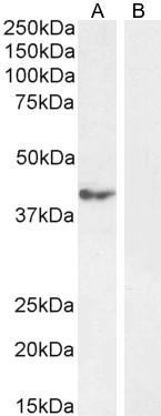
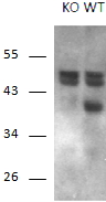
Peptide ELISA: antibody detection limit dilution 1:32000.Western blot: Approx. 40kDa band was observed in Human Cerebellum lysates (calculated MW of 31.9kDa according to NP_001272320.2). This molecular weight is observed by other commercial sources, and was blocked by incubation with the immunizing peptide. Recommended concentration: 1-1.5ug/ml. Primary incubation 1 hour at room temperature. Approx. 40kDa band corresponding to isoform 1L, was observed in Wild-type Mouse Brain lysates, which is not present in the KO mouse. Additional 55kDa bands were consistently observed in both the WT and KO Mouse, and are therefore a non-specific signal. Recommended concentration: 1-3µg/ml. Primary incubation 1 hour at room temperature. Data kindly provided by Dana Crawford, PhD, Albany Medical College, NY, USA.
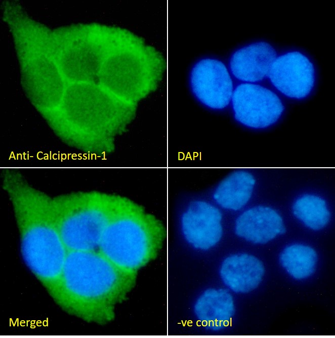
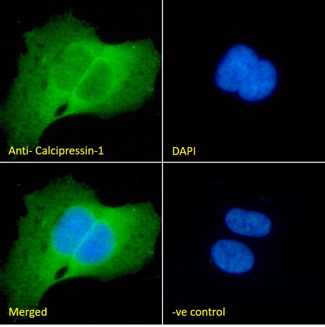
Immunofluorescence: Strong expression of the protein seen in the cytoplasm of A431 and U2OS cells. Recommended concentration: 10µg/ml.
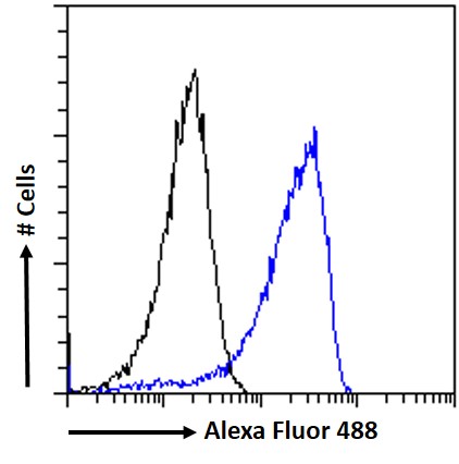
Flow Cytometry: Flow cytometric analysis of A431 cells. Recommended concentration: 10ug/ml.
Species Reactivity
Tested: Human, MouseExpected from sequence similarity: Human, Mouse, Rat, Dog, Cow
Product Reviews
Please login to review this product.- Western blot Guidelines
- Everest Western blot labeling
- Western blot Trouble shooting guide
- IHC staining on paraffin sections
- Immunofluorescence Protocol
- Flow Cytometry Protocol
- MSDS - All Goat antibodies
- Tissue Lysate Preparation
- Cell Lysate Preparation
- Blocking with the immunizing peptide
- LsBio IHC Protocol