
Search
Type in product code or name:
Goat Anti-OGT Antibody (EB07626)
| Code | Name | Applications | Availability | Size | Tested species | Price | Grade | |
|---|---|---|---|---|---|---|---|---|
| EB07626 | Goat Anti-OGT Antibody | Pep-ELISA, WB, IHC, IF, FC | 100µg specific antibody in 200µl | Human, Rat | $464.00 |  |
||
Ordering Information forOrder direct from EverestOrder offline99% of products are held in stock for immediate shipment, see individual product pages for availability. Please order through the US Dollar shopping cart (links on each product page) or we accept orders by email ([email protected])
In stock orders received by 11 am PST on Monday through Friday are shipped the same day. Orders are shipped by our parent company Vector Laboratories, 6737 Mowry Avenue, Newark CA 94560, USA. |
||||||||
Target Protein
Principal Names: OGT, O-linked N-acetylglucosamine (GlcNAc) transferase (UDP-N-acetylglucosamine:polypeptide-N-acetylglucosaminyl transferase), FLJ23071, HRNT1, MGC22921, O-GLCNAC, O-GlcNAc transferase p110 subunit O-linked GlcNAc transferase, uridinediphospho-N-acetylglucosamine:polypeptide beta-N-acetylglucosaminyl transferaseOfficial Symbol: OGT
Accession Number(s): NP_858058.1; NP_858059.1
Human GeneID(s): 8473
Non-Human GeneID(s): 108155 (mouse), 26295 (rat)
Important Comments: This antibody is expected to recognise both reported isoforms (NP_858058.1 and NP_858059.1
Immunogen
Peptide with sequence C-YEHPKDLKLSDGR, from the internal region of the protein sequence according to NP_858058.1; NP_858059.1.
Please note the peptide is available for sale.
Purification and Storage
Purified from goat serum by ammonium sulphate precipitation followed by antigen affinity chromatography using the immunizing peptide.
Supplied at 0.5 mg/ml in Tris saline, 0.02% sodium azide, pH7.3 with 0.5% bovine serum albumin.
Aliquot and store at -20°C. Minimize freezing and thawing.
Applications Tested
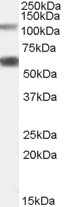
Peptide ELISA: antibody detection limit dilution 1:64000.Western blot: Approx 110kDa band observed in Rat Pancreas lysates calculated MW of 116kDa according to NP_858058.2). An additional band of unknown identity was also consistently observed at 60kDa. This band was successfully blocked by incubation with the immunising peptide. Recommended concentration: 0.05-0.2µg/ml. Primary incubation 1 hour at room temperature. Preliminary testing was unsuccessful on Mouse Brain for this particular batch.
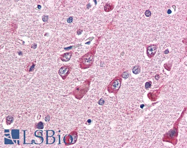
IHC: Paraffin embedded Human Brain (Cortex). Recommended concentration: 5µg/ml.
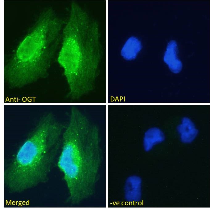
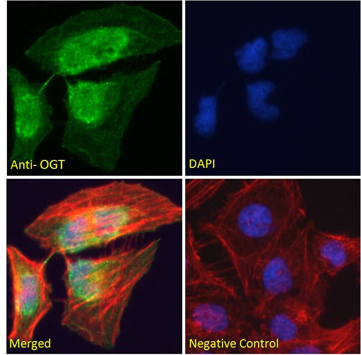
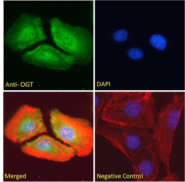
Immunofluorescence: Strong expression of the protein seen in the nucleus of HeLa, U2OS and Glioblastoma U251 cells. Recommended concentration: 10µg/ml.
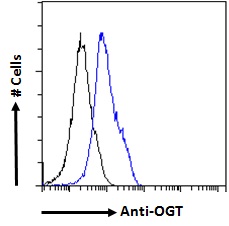
Flow Cytometry: Flow cytometric analysis of HEK293 cells. Recommended concentration: 10ug/ml.
Species Reactivity
Tested: Human, RatExpected from sequence similarity: Human, Mouse, Rat, Dog, Cow
Product Reviews
Please login to review this product.- Western blot Guidelines
- Everest Western blot labeling
- Western blot Trouble shooting guide
- IHC staining on paraffin sections
- Immunofluorescence Protocol
- Flow Cytometry Protocol
- MSDS - All Goat antibodies
- Tissue Lysate Preparation
- Cell Lysate Preparation
- Blocking with the immunizing peptide
- LsBio IHC Protocol