Formulation Supplied at 0.5 mg/ml in Tris saline, 0.02% sodium azide, pH7.3 with 0.5% bovine serum albumin.
| |
Unit Size 100 µg | |
Storage Instructions Aliquot and store at -20°C. Minimize freezing and thawing. | |
Synonym / Alias Names peptidyl-cysteine S-nitrosylase GAPDH|HEL-S-162eP|epididymis secretory sperm binding protein Li 162eP|glyceraldehyde 3-phosphate dehydrogenase|aging-associated gene 9 protein|MGC88685|GAPD|G3PD|HGNC:4141|glyceraldehyde-3-phosphate dehydrogenase|GAPDH | |
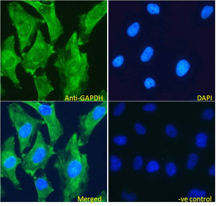
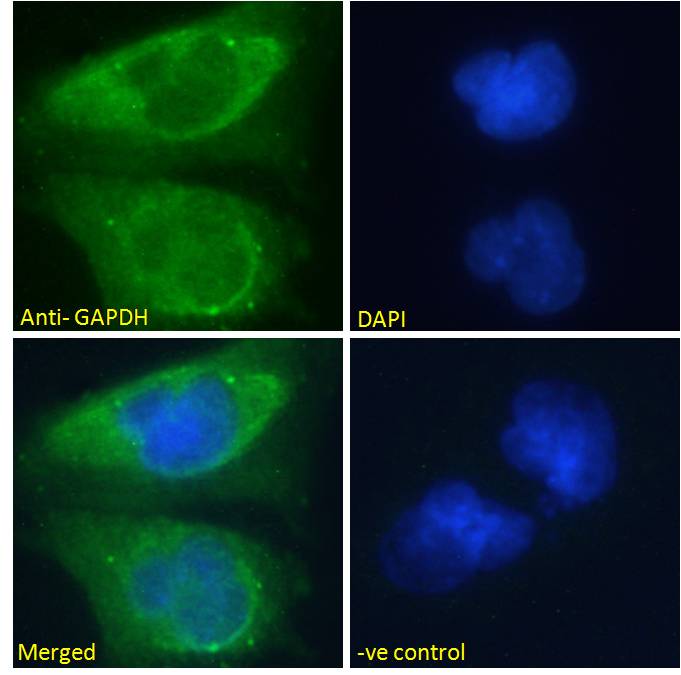
Usage Summary Immunofluorescence: Strong expression of the protein seen in the cytoplasm of U251 and HeLa cells. Recommended concentration: 5-10µg/ml. | |
Accession ID NP_002037.2; NP_001243728.1 | |
Blocking Peptide EBP07069 | |
Immunogen Peptide with sequence C-GVNHEKYDNSLK, from the internal region of the protein sequence according to NP_002037.2; NP_001243728.1. | |
Product Comments This antibody is expected to recognize both reported isoforms (NP_002037.2; NP_001243728.1). Reported variants represent identical protein: NP_001276674.1, NP_002037.2, NP_001276675.1. GAPDH is constitutively expressed in almost all tissues at high levels. It is therefore a useful marker when a loading/positive control is required in western blotting. | |
Peptide Sequence C-GVNHEKYDNSLK | |
Purification Method Purified from goat serum by ammonium sulphate precipitation followed by antigen affinity chromatography using the immunizing peptide. | |
Shipping Instructions Refrigerated | |
Predicted Species Human, Mouse, Rat, Dog | |
Reactive Species Human, Mouse, Rat | |
Human Gene ID 2597 | |
Mouse Gene ID 14433 | |
Rat Gene ID 24383 | |
Product Grade  | |
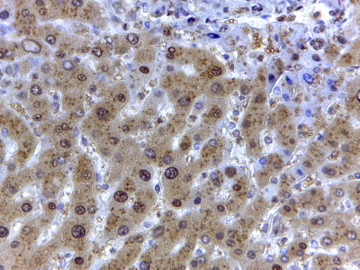
IHC Results In paraffin embedded Human Liver shows textured cytoplasm staining in hepatocytes. Recommended concentration: 2µg/ml. | |
ELISA Detection Limit Antibody detection limit dilution 1:16000. | |
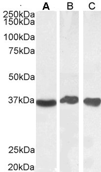
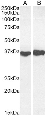
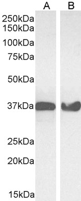
Western Blot Approx 37kDa band observed in Human Liver, Testis and Tonsil, and in Mouse Liver and Rat Heart lysates, and in cell lysates of HeLa and NIH3T3 (calculated MW of 36.1kDa according to Human NP_002037.2, Mouse NP_032110.1, and Rat NP_058704.1). Recommended concentration: 0.03-0.1µg/ml. Primary incubation 1 hour at room temperature. This product has been successfully used in WB on Human: PMID: 29953622, PMID: 29158223 and https://doi.org/10.1101/2022.01.21.477266, and in WB on Rat: PMID: 31087788 and 34584880. | |
Application Type Pep-ELISA, WB, IHC, IF |
Goat Anti-GAPDH (Internal) Antibody
$431.00
| SKU | Unit Size | Price |
|---|---|---|
Select a unit size:
Selected References [{"pmid": 29158223, "intro": "This antibody has been successfully used in Western blot on Human:", "title": "TGF-? induces oncofetal fibronectin that, in turn, modulates TGF-? superfamily signaling in endothelial cells", "author": "Elisa Ventura, Michael Weller, Will Macnair, Katja Eschbach, Christian Beisel, Cinzia Cordazzo, Manfred Claassen, Luciano Zardi and Isabel Burghardt", "journal": "J Cell Sci. 2018 Jan 9;131(1)."}, {"pmid": 21738400, "intro": "This antibody (previous batch) has been successfully used in Western blot on Human:", "title": "The expression of retinal cell markers in human retinal pigment epithelial cells and their augmentation by the synthetic retinoid fenretinide.", "author": "Carr AJ, Vugler AA, Yu L, Semo M, Coffey P, Moss SE, Greenwood J.", "journal": "Mol Vis. 2011;17:1701-15."}, {"pmid": 19204785, "intro": "This antibody (previous batch) has been successfully used in Western blot on Human:", "title": "Molecular characterization and functional analysis of phagocytosis by human embryonic stem cell-derived RPE cells using a novel human retinal assay.", "author": "Carr AJ, Vugler A, Lawrence J, Chen LL, Ahmado A, Chen FK, Semo M, Gias C, da Cruz L, Moore HD, Walsh J, Coffey PJ.", "journal": "Mol Vis. 2009;15:283-95."}, {"pmid": 18926821, "intro": "This antibody (previous batch) has been successfully used in the following paper:", "title": "Elucidating the phenomenon of HESC-derived RPE: anatomy of cell genesis, expansion and\r\nretinal transplantation.", "author": "Vugler A, Carr AJ, Lawrence J, Chen LL, Burrell K, Wright A, Lundh P, Semo M, Ahmado A, Gias C, da Cruz L, Moore H, Andrews P, Walsh J, Coffey P.", "journal": "Exp Neurol. 2008 Dec;214(2):347-61."}, {"pmid": 29953622, "intro": "This antibody has been successfully used in Western blot on Human:", "title": "Epidermal growth factor receptor and ligand family expression and activity in glioblastoma.", "author": "von Achenbach C, Weller M, Szabo E.", "journal": "J Neurochem. 2018 Jun 28."}, {"pmid": 29480969, "intro": "This antibody has been successfully used in the following paper:", "title": "Transcriptional control of O6 -methylguanine DNA methyltransferase expression and temozolomide resistance in glioblastoma.", "author": "Happold C, Stojcheva N, Silginer M, Weiss T, Roth P, Reifenberger G, Weller M.", "journal": "J Neurochem. 2018 Mar;144(6):780-790"}, {"pmid": 31087788, "intro": "This antibody has been successfully used in Western blot on Rat:", "title": "miR?455?5p Overexpression Reduces Rat Lung Alveolar Type II Cell Proliferation by Downregulating STRA6", "author": "Zheng J, He Q, Tang H, Xia H", "journal": "Anat Rec (Hoboken). 2019 May 14."}, {"pmid": 30081309, "intro": "This antibody (previous batch) has been successfully used in Western blot on Human:", "title": "Equine MX2 is a restriction factor of equine infectious anemia virus (EIAV)", "author": "Meier K, Jaguva Vasudevan AA, Zhang Z, Bähr A, Kochs G, Häussinger D, Münk C", "journal": "Virology. 2018 Oct;523:52-63. doi: 10.1016/j.virol.2018.07.024"}, {"pmid": 32371586, "intro": "This antibody has been successfully used in the following paper:", "title": "Depatuxizumab mafodotin (ABT-414)-induced glioblastoma cell death requires EGFR overexpression, but not EGFRY1068 phosphorylation.", "author": "von Achenbach C, Silginer M, Blot V, Weiss WA, Weller M", "journal": "Mol Cancer Ther. 2020 May 5. pii: molcanther.0609.2019."}, {"pmid": 34584880, "intro": "This antibody has been successfully used in Western blot on Rat:", "title": "miR-363-3p inhibits rat lung alveolar type II cell proliferation by downregulating STRA6 expression and induces cell apoptosis via cellular oxidative stress and G1-phase cell cycle arrest", "author": "Jintao Zheng, Shibo Zhu, Huiyu Xu, Jiequan Li, Huajian Tang, Yanfen Zhou, Zhaomei Huang, Guoqing Liu", "journal": "Transl Pediatr. 2021 Aug; 10(8): 2095–2105."}, {"pmid": 0, "intro": "This antibody has been successfully used in Western blot on Human:", "title": "EFA6R suppresses ovarian cancer cell migration and invasion", "author": "Salman Tamaddon-Jahromi, Kate Murphy, William Walker and Venkateswarlu Kanamarlapudi", "journal": "https://doi.org/10.1101/2022.01.21.477266"}] |
Documents |